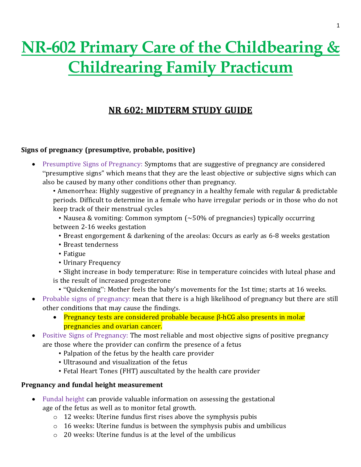
Signs of pregnancy
presumptive (subjective signs) Amenorrhea, nausea, vomiting, increased urinary frequency,
excessive fatigue, breast tenderness, quickening at 18–20 weeks
probable (objective signs) Goodell sign (sof
...
Signs of pregnancy
presumptive (subjective signs) Amenorrhea, nausea, vomiting, increased urinary frequency,
excessive fatigue, breast tenderness, quickening at 18–20 weeks
probable (objective signs) Goodell sign (softening of cervix)
Chadwick sign (cervix is blue/purple)
Hegar’s sign (softening of lower uterine segment)
Uterine enlargement
Braxton Hicks contractions (may be palpated by 28 weeks)
Uterine soufflé (soft blowing sound due to blood pulsating through the placenta)
Integumentary pigment changes
Ballottement, fetal outline definable, positive pregnancy test (could be hydatidiform mole,
choriocarcinoma, increased pituitary gonadotropins at menopause)
positive (diagnostic signs) Fetal heart rate auscultated by fetoscope at 17–20 weeks or by Doppler at
10–12 weeks
Palpable fetal outline and fetal movement after 20 weeks
Visualization of fetus with cardiac activity by ultrasound (fetal parts visible by 8 weeks)
Pregnancy and fundal height measurement
Signs of pregnancy (presumptive, probable, positive)
Pregnancy and fundal height measurement As pregnancy progresses, the
fundus rises out of the pelvis (Figure 29-1). At 12 weeks’ gestation, the fundus is
located at the level of the symphysis pubis. By week 16, it rises to midway between
symphysis pubis and the umbilicus. By 20 weeks’ gestation, the fundus is typically at the
same height as the umbilicus. Until term, the fundus enlarges approximately 1 cm per
week. As the time for birth approaches, the fundal height drops slightly. This process,
which is commonly called lightening, occurs for a woman who is a primigravida around
38 weeks’ gestation but may not occur for the woman who is a multigravida until she
goes into labor
Naegele’s rule
Add seven days to the first day of your LMP and then subtract three months. For
example, if your LMP was November 1, 2017: Add seven days (November 8, 2017).
Subtract three months (August 8, 2017).
The EDD is calculated by adding seven days to the first day of the last menstrual period, subtracting
three months and adding one year.
This formula is known as Naegele's Rule. For example, if the patient's last menstrual period, LMP,
was on August 10, 2019, the EDD would be calculated as follows. LMP equals August 10, 2019 plus
seven days. August 17, 2019, minus three months. May 17, 2019 plus one year and that equals May
17, 2020.
Hematological changes during pregnancy
During pregnancy, the heart is displaced upward and to the left within the chest cavity
by the gravid uterus’s pressure on the diaphragm. As pregnancy progresses, the risk for
inferior vena cava and aortic compression leading to supine hypotension increases
when the woman lies in a supine position. To avoid hypotension and potential syncope,
the woman should be advised to lie in a left lateral position. Hemodynamic changes and
anatomic changes also may alter vital signs in the pregnant woman (Table 29-2).
Cardiac output in pregnancy increases by 30% to 50% over that in women who are not
pregnant (Blackburn, 2013; Ouziunian & Elkayam, 2012). This increase
peaks in the early third trimester and is maintained until birth. Half of the total increase
in cardiac output, however, occurs by the eighth week of pregnancy (Blackburn,
2013). Therefore, women with cardiac disease may become symptomatic during the
first trimester. Stroke volume is also increased during pregnancy by 20% to 30%. These
increases in cardiac output and stroke volume allow for the 30% increase in oxygen
consumption observed during pregnancy.
TABLE 29-2 Vital Sign Changes in Pregnancy
Vital Sign Changes in Pregnancy Measurement Alterations in
Pregnancy
Heart rate
and heart
sounds
Volume of the first heart sound
may be increased with splitting.
Third heart sound may be
detected.
Systolic murmurs may be detected.
Increases by 15–20 beats/min by
32 weeks’ gestation.
Palpate the maternal pulse when
auscultating the fetal heart rate to
be able to distinguish between the
two.
Respiratory
rate
Increases by 1–2 breaths/min None
BP First trimester: same as
prepregnancy values
Second trimester: systolic BP
decreases by 2–8 mm Hg and
diastolic BP decreases by 5–15 mm
Hg due to peripheral vascular
resistance
Third trimester: gradually returns to
prepregnancy values
Use of an automated cuff may
improve accuracy of
measurement, as some pregnant
women do not have a fifth
Korotkoff sound.
Systolic and diastolic BP may be
16 mm Hg higher when taken
while the woman is sitting.
BP readings may decrease in the
maternal left lateral position.
Abbreviation: BP, blood pressure.
Data from Jarvis, C. (2016). Physical examination and health assessment (7th ed.). St. Louis, MO:
Saunders Elsevier; Ouziunian, J., & Elkayam, U. (2012). Physiologic changes during normal
pregnancy and delivery. Cardiology Clinics, 30, 317–329; Tan, E., & Tan, E. (2013). Alterations in
physiology and anatomy during pregnancy. Best Practice & Research Clinical Obstetrics &
Gynaecology, 27, 791–802.
During pregnancy, blood volume increases by 30% to 50%, or 1,100 to 1,600 mL
(Ouziunian & Elkayam, 2012), and peaks at 30 to 34 weeks’ gestation. The
increase in blood volume improves blood flow to the vital organs and protects against
excessive blood loss during birth. Fetal growth during pregnancy and newborn weight
are correlated with the degree of blood volume expansion.
Of the blood volume expansion occurring during pregnancy, 75% is considered to be
plasma (King et al., 2015). There is also a slight increase in red blood cell volume
(RBC). The blood volume changes result in hemodilution, which leads to a state of
physiologic anemia during pregnancy. As the RBC volume increases, iron demands also
increase. Leukocytosis occurs in pregnancy, with white blood cell counts increasing to
as much as 14,000 to 17,000 cells per mm3 of blood (Table 29-3). Clotting factors
increase as well, creating a risk for clotting events during pregnancy.
Systemic vascular resistance is reduced due to the effects of progesterone,
prostaglandins, estrogen, and prolactin. This lowered systemic vascular resistance, in
combination with inferior vena cava compression, is partly responsible for the
dependent edema that occurs in pregnancy. Epulis of pregnancy, or hypertrophy of the
gums accompanied by bleeding, may also occur and is due to decreased vascular
resistance and increase in the growth of capillaries during pregnancy (Jarvis, 2016).
Indications and contraindications for prescribing combined estrogen
vs. progesterone-only birth control
Progestin-only contraceptives are used continuously; there is no hormone-free interval,
as occurs with combined methods. These contraceptive methods have minimal effects
on coagulation factors, blood pressure, or lipid levels and are generally considered safer
for women who have contraindications to estrogen, such as cardiovascular risk factors,
migraine with aura, or a history of VTE. In spite of this belief, the product labeling for
some progestin-only products mimics the labeling for products containing estrogen.
The U.S. Medical Eligibility Criteria for Contraceptive Use (CDC, 2010;
see Appendix 11-A) can be used to identify appropriate candidates for progestinonly contraception.
Progestin-only contraceptives do not provide the same cycle control as methods
containing estrogen, and unscheduled bleeding is common with all progestin-only
methods. Typically, unscheduled bleeding occurs most frequently during the first 6
months of method use, with a substantial number of users becoming amenorrheic by 12
months of use (Hubacher, Lopez, Steiner, & Dorflinger, 2009). Overall blood
loss decreases over time, making progestin-only methods protective against irondeficiency anemia. With appropriate counseling, many women see amenorrhea as a
benefit of these methods.
All progestin-only methods are likely to improve menstrual symptoms, including
dysmenorrhea, menorrhagia, premenstrual syndrome, and anemia (Burke, 2011).
The thickening of cervical mucus seen with progestin methods is protective against PID.
Progestin-only contraceptives include the progestin-only pill (POP), an injection, an
implant, and three progestin-containing intrauterine devices. The implant and devices
are covered in the section on long-acting reversible contraception.
The U.S. Medical Eligibility Criteria for Contraceptive Use (CDC, 2010) is a
comprehensive, evidence-based guide for determining whether women have relative or
absolute contraindications to contraceptive methods. The Medical Eligibility
Criteria uses the following four classification categories of whether a person can use or
should not use a method:
Category 1: a condition for which there is no restriction for the use of the
contraceptive method
Category 2: a condition where the advantages of using the method generally
outweigh the theoretical or proven risks
Category 3: a condition where the theoretical or proven risks usually outweigh
the advantages of using the method
Category 4: a condition that represents an unacceptable health risk if the
contraceptive method is used
Menstrual cycle physiology
The initiation of menstruation, called menarche, usually happens between the ages of
12 and 15. Menstrual cycles typically continue to age 45 to 55, when menopause
occurs. Many women find themselves reluctant to discuss the existence and normality
of menstruation. The word menstruation has been replaced by a variety of euphemisms,
such as the curse, my period, my monthly, my friend, the red flag, or on the rag.
Most women experience deviations from the average menstrual cycle during their
reproductive years. As a result, it is not uncommon for women to display certain
preoccupations regarding their menstrual bleeding, not only in relation to the regularity
of its occurrence, but also in regard to the characteristics of the flow, such as volume,
duration, and associated signs and symptoms. Unfortunately, society has encouraged
the notion that a woman’s normalcy is based on her ability to bear children. This
misperception has understandably forced women to worry over the most miniscule
changes in their menstrual cycles. Indeed, changes in menstruation are one of the most
frequent reasons why women visit their clinician.
Numerous patterns in the secretion of estrogens and progesterone are possible; in fact,
it is difficult to find two cycles that are exactly the same. Studies that include women of
different ethnicities, occupations, genetics, nutritional status, and age have
demonstrated that the length and duration of the menstrual cycle vary widely (Assadi,
2013; Johnson et al., 2013; Karapanou & Papadimitriou, 2010).
Menarche is the most readily evident external event that indicates the end of one
developmental stage and the beginning of a new one. It is now believed that body
composition is critically important in determining the onset of puberty and menstruation
in young women (Ferin & Lobo, 2012). The ratio of total body weight to lean body
weight is probably the most relevant factor, and individuals who are moderately obese
(i.e., 20–30% above their ideal body weight) tend to have an earlier onset of menarche
(Johnson et al., 2013). Widely accepted standards for distinguishing what are
regular versus irregular menses, or normal versus abnormal menses, are generally
based on what is considered average and not necessarily typical for every woman.
According to these standards, the normal menstrual cycle is 21 to 35 days with a
menstrual flow lasting 4 to 6 days, although a flow for as few as 2 days or as many as 8
days is still considered normal (Ferin & Lobo, 2012).
The amount of menstrual flow varies, with the average being 50 mL; nevertheless, this
volume may be as little as 20 mL or as much as 80 mL. Generally, women are not
aware that anovulatory cycles and abnormal uterine bleeding (changes in bleeding
outside of normal; see Chapter 24) are common after menarche and just prior to
menopause (Ferin & Lobo, 2012; Fritz & Speroff, 2011). Menstrual cycles
that occur during the first 1 to 1.5 years after menarche are frequently irregular due to
the immaturity of the hypothalamic–pituitary–ovarian axis (Fritz & Speroff, 2011).
Vaccines during pregnancy
Live vaccines are contraindicated during pregnancy (MMR, Oral Polio, Varicella
& FluMist)
Injectable influenza vaccine is an inactivated virus and is safe to use in
pregnancy
Ask if the woman has ever known anyone with tuberculosis or traveled to areas where
tuberculosis is common. If she is at risk, she should receive a tuberculin skin test when
she can return in 48 to 72 hours. Past history of varicella is important, as well as the
woman’s vaccine history, to determine if she is at risk for chickenpox.
Women can receive vaccines in pregnancy (Table 30-1). The Centers for Disease
Control and Prevention (CDC) updates the adult vaccine schedule often, and this
information can be easily accessed on its website. The CDC website also includes
detailed information about safety of vaccines for travel of local disease outbreaks during
pregnancy (CDC, 2014). All women who are pregnant should be offered the influenza
vaccine during flu season, though live attenuated influenza vaccine (LAIV [FluMist])
should not be given to pregnant women. All women should be encouraged to receive a
tetanus, diphtheria, and acellular pertussis (Tdap) vaccination in the third trimester
(CDC, 2016). Other vaccines, such as hepatitis B, can be administered if the woman
is at risk (CDC, 2016).
During pregnancy, women have a decreased immune response to pathogens, making
them more susceptible to infection. If a woman has cats, she should be careful to avoid
contracting toxoplasmosis—an infection that is spread through cat feces. Someone else
should change the cat litter box daily to prevent contact with the Toxoplasma
gondii parasite. Wearing gloves while gardening, and careful hand washing are also
essential. More information and patient handouts are available for free at the CDC
website.
TABLE 30-1 Vaccines in Pregnancy
Recommended Each Preg
nancy
Rationale Timing
Influenza (flu)a Women who are pregnant are at
increased risk for flu-related
complications.
Any
gestation
when the
injection is
available
[Show More]


![Preview of WEEK **5 [Chamberlain university]NR 602 IHUMAN Case Week #5 Deborah Arnaudin 54 Year Old](https://browseimages.nyc3.digitaloceanspaces.com/paper-images/2026/03/31/zbQKePwt2026-03-31-11-4669cc328fcad96.png)