Week 7: Behavioral, Neurologic, and Digestive Disorders - Discussion Part One
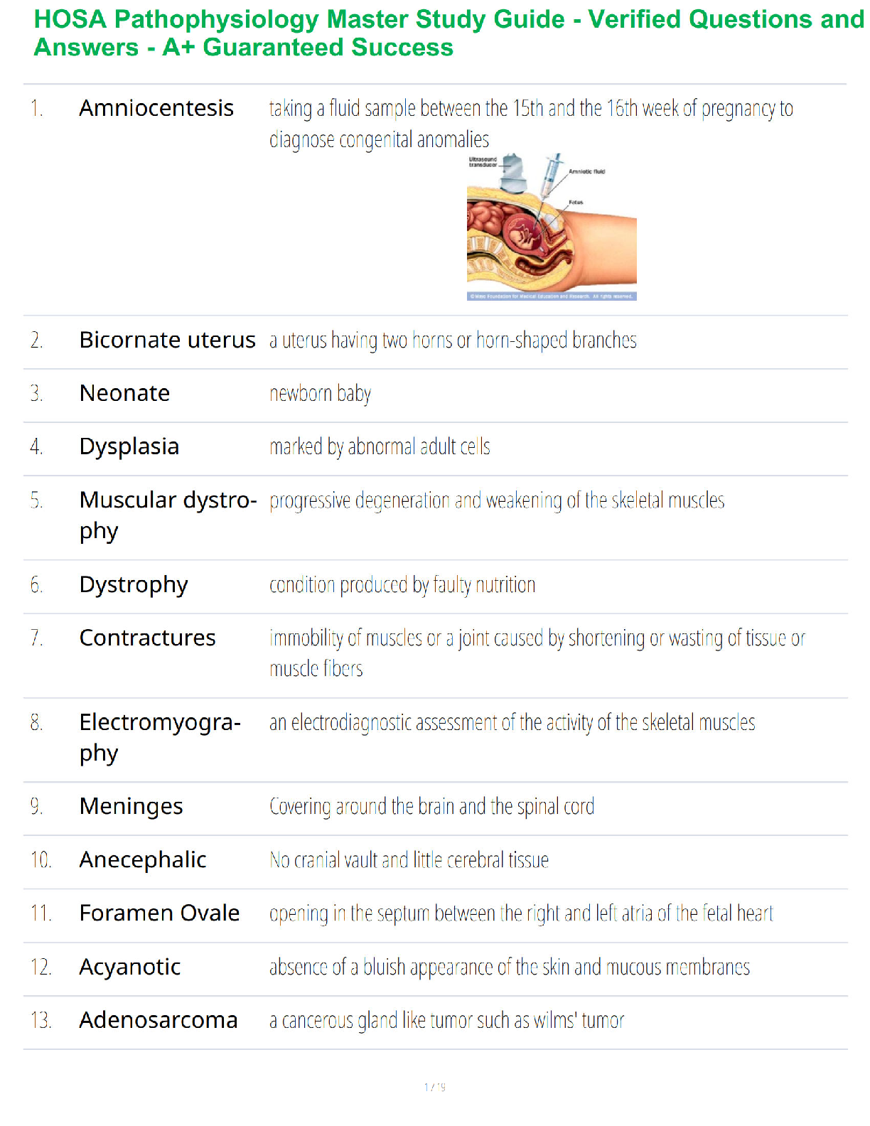
Loading... Discussion This week's graded topics relate to the following Course Outcomes (COs).
1 2 3 4 5 6 7
Analyze pathophysiologic mecha
...
Week 7: Behavioral, Neurologic, and Digestive Disorders - Discussion Part One
Loading... Discussion This week's graded topics relate to the following Course Outcomes (COs).
1 2 3 4 5 6 7
Analyze pathophysiologic mechanisms associated with selected disease states. (PO 1)
Differentiate the epidemiology, etiology, developmental considerations, pathogenesis, and clinical and laboratory manifestations of specific disease processes. (PO 1)
Examine the way in which homeostatic, adaptive, and compensatory physiological mechanisms can be supported and/or altered through specific therapeutic interventions. (PO 1, 7)
Distinguish risk factors associated with selected disease states. (PO 1)
Describe outcomes of disruptive or alterations in specific physiologic processes. (PO 1)
Distinguish risk factors associated with selected disease states. (PO 1)
Explore age-specific and developmental alterations in physiologic and disease states. (PO 1, 4)
Discussion Part One (graded)
You are at the local mall and you see a patient who appears to be homeless by his physical appearance and you witness the person “walk 50 feet to a table sit down, and after 5 seconds he gets up and walks to a tree and urinates on it” He repeats this action 5 times apparently oblivious to his surroundings. When the police come he ignores them as if they aren’t there. Later, you go to work and sitting in exam room 3 is the same person! Now, he is your patient, when you talk to him he has no recollection of his behavior by the mall.• What is your differential diagnosis?
• What tests do you order?
• An MRI comes back and there seems to be a lesion in the temporal lobe does this change
your differential? The EEG also comes back with unusual excitatory activity. What is your
definitive diagnosis? In retrospect did anything bias your first differential?
Responses
Rechel DelAntar
Differential Diagnosis
Hello professor and Class,
Differential Diagnosis
This is a case of a patient who was previously seen in the mall looking homeless in physical
appearance who exhibits repetitive movement such as “walking to a table, sitting up, walking to a tree
and urinating” oblivious to the people around him and his surroundings. Upon initial exam he has no
recollections of previous events. Based on this history, we may consider:
Seizures = specifically Complex Focal seizure (temporal or psychomotor seizure)with a simple partial
onset followed by impairment of consciousness. In this type of seizure, the patient is able to interact
with the environment with a purposeful, although inappropriate movement. Most characteristic event of
this type of seizure is the automatism; common examples of automatisms are lip smacking, chewing,
facial grimacing, swallowing movements, and patting, picking, or rubbing oneself or one’s clothing. The
body may stiffen but the patient will continue to perform complex activities of which they are involved in
such as driving. Witnesses may not recognize that anything is wrong. Temporal lobe seizures generally
last 11 seconds to 8 minutes (average 2 minutes) and are followed by several minutes of postictal
confusion (McCance, K.L., 2013).
Diagnostic testing = Laboratory studies have to be done to rule out potential causes or triggers to
seizures. MRI of the brain can be performed to check if structural lesions are causing the seizure event
and is helpful in assessing temporal seizures. Temporal lobe seizures commonly result from damage to
specific areas in that part of the brain. This can be due to a head injury, infection, or damage to a portion
of the temporal lobe due to lack of oxygen, brain tumors, genetic syndromes, or lesions of any sort.
Many of these problems also produce brain-tissue scarring called mesial temporal sclerosis. EEG within
24 hours is more sensitive for diagnosing epileptiform abnormalities as it is able to localize seizure focus.
Brain cells communicate with each other and produce our consciousness, thoughts, and actions by
electrochemical processes. Certain patterns of electrical activity disrupt this normal function of the brain
and spread in abnormal patterns within the brain. This process can be seen on an EEG (Walter, B.,
2013).
With the patient’s MRI results coming back with temporal lobe lesion and an EEG reading of
unusual excitatory activity, my differential diagnosis remains the same. The results support the
diagnosis. In retrospect, the patient’s inability to recollect his actions made me think that he was not in
control of his actions and also reminded me of post ictal stage of seizure when patients were unable to
recall the seizure event is what gave bias to my differential diagnosis.
References:
McCance, K. L., Huether, S. E., Brashers, V. L., & Rote, N. S. (2013). Pathophysiology: The
biologic basis
for disease in adults and children (7th ed.). St. Louis, MO: Mosby.
Walter, B. (2013). Bradley’s neurology in clinical practice (6 ed.). Philadelphia, PA: Elsevier,
Saunders.
th
Instructor Brown reply to Rechel DelAntar
RE: Differential Diagnosis
What symptoms are possible with right vs. left side lesion?
6/14/2016 2:23:24 PM
6/12/2016 12:44:25 PMRechel DelAntar reply to Instructor Brown
RE: Differential Diagnosis
6/14/2016 10:20:29 PM
Hello Professor and Class,
Right side vs. Left side lesion
The brain is the control center for all human activity, including vital processes
(breathing and moving) as well as thinking, judgment, and emotional reactions. Brain lesions
can be caused by injury, infection, exposure to certain chemicals, problems with the immune
system, and more. Typically, their cause is unknown. Symptoms experienced by the patient
vary depending on the location, type and size of the lesion. The brain is divided into two halves
(hemispheres). The left half controls movement and sensation in the right side of the body, and
the right half controls movement and sensation in the left side. Therefore, damage to the right
side of the brain may cause movement problems or weakness on the body's left side. For
majority of the population, the left half of the brain is responsible for verbal and logical functions
including language (listening, reading, speaking, and writing), thought and memory involving
words. Patients with this type of lesion will exhibit right side weakness, aphasia, slow speech and
decreased attention span. The right half is responsible for nonverbal and intuitive functions such
as putting bits of information together to make up an entire picture, recognizing oral and visual
patterns and designs (music and art), and expressing and understanding emotions. Patients
with this type of lesion will exhibit left sided weakness, will have difficulty with complex
communication such as difficulty identifying relevant information, inability to interpret body
language and relevant information. They tend to be very literal in their interpretation of things
and situations. Left hemispheric damage may produce a right hemianopsia or quadranopsia.
They have flat affect and at times are impulsive (Novack, T., n.d.). Right Hemispheric damage
may not only produce a left homonymous hemianopsia or quadranopsia, but it may also produce
a severe attention disorder to the left side called left hemispatial inattention or visual neglect.
Spatial orientation, body position and nonverbal communications may become impaired in some
individuals. Emotional and behavioral problems may occur. Thinking skills may be effected.
Meanwhile, many patients will be unaware of the full extent of their impairment. They may even
deny they have a problem (Manasco, H., 2014).
References:
Manasco, H. (2014). Introduction to neurogenic Communication Disorders.
Burlington, MA: Jones and Bartlett Learning.
Novack, T. (n.d.). Understanding TRI part 2: Brain Injury Impact on Individual
Functioning. Retrieved from http://www.msktc.org/tbi/factsheets/
Understanding-TBI/Brain-Injury-Impact-On-
Individuals-Functioning.
Brittany Heller
AD
6/12/2016 1:51:25 PM
"Alzheimer disease is a neurodegenerative disorder of uncertain cause and pathogenesis that
primarily affects older adults and is the most common cause of dementia" ( Wolk & Dickerson, 2016, p.
1). AD typically affects patients older than 60 years and is rarely occurs in less than 60 (Wolk &
Dickerson, 2016, p. 1). Some have suggested that there is a mutation in genes that alter beta-amyloid
protein production and metabolism (Wolk & Dickerson, 2016, p. 1). Genes that are mutated include
APP, PSEN1, and PSEN2 (Wolk & Dickerson, 2016, p. 1). AD also had three common areas of cortical
thinning patterns that were observed which are the medial temporal, diffuse, and patietal dominant
atrophy subtypes (Hwang, Kim, Jeon, Lee, HOne, Roh, Lee, Koh, & Noh, 2016, p. 1). The thinning of the
cortical areas may suggest a predictive pattern in the pathophysiology of AD. Memory impairment is the
most common symptom of AD. "Executive dysfunction and visuospatial impairment are often presentrelatively early, while deficits in language and behavioral symptoms often manifest late in the disease
course" (Wolk & Dickerson, 2016, p. 2). Other symptoms can include executive function and
judgement/problem solving, behavioral and psychological symptoms, apraxia, olfactory dysfunction,
sleep disturbances, and seizures ( Wolk & Dickerson, 2016, p. 5).
After completing a standardized mental status scale, the first test I would order would be an MRI. "Brain
MRI can document potential alternative or additional diagnoses of cerebrovascular disease, other
structural disease, and regional brain atrophy suggesting frontotemporal dementia or other types of
neurodegenerative disease" ( Wolk & Dickerson, 2016, p. 6).
If the MRI came back with a lesion in the temporal lobe, this would not change my diagnosis. "The
most characteristic focal finding in AD is reduced hippocampal volume and medial temporal lobe atrophy"
(Wolk & Dickerson, 2016, p. 6). Seizures are one of the clinical manifestations that can be present in
AD. "Temporal lobe epilepsy is the most common form of partial or localization related to epilepsy"
(Holmes, Sirven, & Fisher, 2013, p. 1). For temporal lobe seizures, an EEG would be essential for
diagnosing purposes. On an EEG, sharp waves or spikes would be seen showing unusual activity
(Holmes et al., 2013, p. 4).
After thinking about this diagnosis, the memory loss was a key factor in deciding on the AD diagnosis.
Due to his repetitive actions, I thought he could possibly have OCD as well. Not having the age of the
patient also made this difficult to diagnosis this. Not recognizing the police also had a judgement
impairment which leaned more towards AD as well.
Holmes, G., Sirven, J., & Fisher, R. (2013). What is temporal lobe epilepsy? Epilepsy Foundation.
Retrieved at: http://www.epilepsy.com/learn/types-epilepsy-syndromes/temporal-lobe-epilepsy
Hwang, J., Kim, C., Jeon, S., Lee, J., Hong, Y., Roh, J., Lee, J., Koh, J., 7 Na, D. (2016) Prediction of
Alzheimer's disease pathophysiology based on cortical thickness patterns. Alzheimers Dementia, 2, 58-
67. doi: 10.1016/j.dadm.2015.11.008. Retrieved at: http://www.ncbi.nlm.nih.gov/pubmed/27239533
Wolk, D., & Dickerson, B. (2016). Clinical features and diagnosis of Alzheimer Disease. UpToDate.com.
Retrieved at: http://www.uptodate.com/contents/clinical-features-and-diagnosis-of-Alzheimer-disease?
topickey
Lanre Abawonse reply to Brittany Heller
RE: AD
6/14/2016 8:46:21 PM
Brittany, your narration is great. I would like to add some few points to your diagnosis
and the findings on the magnetic resonance imaging MRI. Just as you selected Alzheimer’s
disease as your number diagnosis, I did the same on my selection. Alzheimer’s disease is one
of many conditions that are worth putting into consideration when patients have results that
indicate temporal lobe lesions. The consideration of symptoms can also include alcoholism and
substance abuse etc. Upon further study of temporal lobe lesions resulting effects on patients,
it has been suggested that there are a behavioral changes in these patients. Gudmundsson et.
al., (2015) suggested that cortical atrophy, indicating neurodegeneration, is commonly seen on
imaging of the aging brain of a patient who has temporal lobe lesions on MRI. In addition,
cortical atrophy may also be an expression of small vessel disease. Prospective population-
based studies using magnetic resonance imaging (MRI) report that white matter changesincrease the risk of subsequent dementia. With these findings, it could suggest that this patient
is going through behavioral changes and this is evidenced by the patient’s lack of insight along
with memory impairment.
Gudmundsson, P., Olesen, P. J., Simoni, M., Pantoni, L., Östling, S., Kern, S., & ... Skoog, I.
(2015). White matter lesions and temporal lobe atrophy related to incidence of both
dementia and major depression in 70-year-olds followed over 10 years. European
Journal Of Neurology, 22(5), 781-e50.
Jonathan Bidey reply to Brittany Heller
RE: AD
Brittany,
Excellent post! You did a wonderful job describing which genes are involved with
Alzheimer’s disease (AD). You also did a wonderful job describing the physical changes
which occur in the brain during AD. I particularly enjoyed your description of the use of
MRI in diagnosing AD. You mentioned hippocampal volume and its correlation to AD.
MRIs are not only used to evaluate hippocampal findings, but also to assess for other
potential causes of symptoms (Li et al., 2014). Studies are suggesting that changes in
hippocampal volume may soon be able to be detected before any symptoms or memory
loss has occurred. By assessing for these changes early on, prior to the onset of disease,
it is possible that therapies will be designed to slow the onset for those at risk (Li et al.,
2014). Excellent post!
-Jonathan
Reference:
Li, M., Oishi, K., He, X., Qin, Y., Gao, F., & Mori, S. (2014). An efficient
approach for differentiating Alzheimer's disease from normal elderly based
on multicenter MRI using gray-level invariant features. PLoS ONE, 9(8),
1-13. http://dx.doi.org/10.1371/journal.pone.0105563
Lorna Durfee
Discussion Part One
Scenario:
You are at the local mall and you see a patient who appears to be homeless by his physical
appearance, and you witness the person “walk 50 feet to a table sit down, and after 5 seconds he
gets up and walks to a tree and urinates on it” He repeats this action 5 times apparently oblivious
to his surroundings. When the police come, he ignores them as if they aren’t there. Later, you go
to work and sitting in exam room 3 is the same person! Now, he is your patient, when you talk to
him he has no recollection of his behavior by the mall.
Repetitive urination, agnosia, aphasia, apraxia, lesion in temporal lobe?
What is your differential diagnosis?
• What tests do you order?
• An MRI comes back, and there seems to be a lesion in the temporal lobe does this change
your differential? The EEG also comes back with unusual excitatory activity. What is your
definitive diagnosis? In retrospect did anything bias your first differential?
Doctor Brown:
DIFFERENTIALS:
6/13/2016 6:35:51 AM
6/17/2016 12:20:14 PMDifferential #1: Head trauma, with brain injury, traumatic brain injury with cognitive dysfunction
Differential #2: Brain dysfunction.
Differential #3: Excessive alcohol consumption and Wernicke-Korsakoff Syndrome
Differential #4: Schizophrenia with agnosia.
Differential #5: Temporal lobe epilepsy.
My chosen differential diagnosis is Differential #1: Head Trauma or traumatic brain injury.
Tests to Order: Mott, McConnon and Rieger (2012) state that guidelines issued by the United
States Department of Veteran Affairs and the United States Department of Defense outline the
management of subacute to chronic MTBI (mild traumatic brain injury). Since patients can present
with varying symptoms, focusing on the symptoms and physical examination findings is the best
course of action (Mott, McConnon, & Rieger, 2012, p. 1047). Along with a physical exam, there
should be a neurologic examination that focuses on mental status, the cranial nerves, deep tendon
reflexes, gross sensation, stability, and strength. Visual fields and acuity need to be tested along
with monitoring of eye movements.
A musculoskeletal exam should also be performed (Mott, McConnon, & Rieger, 2012, p. 1047).
Since there is no previous work up there should be consultation with a subspecialist. A complete
blood count, electrolyte, and TSH. The use of computed tomography and magnetic resonance
imaging of the brain can be useful in this case (Mott, McConnon, & Rieger, 2012, p. 1048). It
would be wise to do neuropsychologic testing as well as testing for memory, attention span, and
visual and spatial coordination. Further referrals to a specialist and rehabilitation specialist would
be pertinent (Mott, McConnon, & Rieger, 2012, p. 1048). A specialized multidisciplinary approach
should be included in our plan of attack (Mott, McConnon, & Rieger, 2012, p. 1050).
An MRI comes back, and there seems to be a lesion in the temporal lobe does this change your
differential? The EEG also comes back with unusual excitatory activity. What is your definitive
diagnosis? In retrospect did anything bias your first differential?
No, my differential does not change. The diagnosis is traumatic brain injury based on the
information presented. From the beginning of this case, I felt that this patient (as a homeless
person) could experience situations that are susceptible to the harsh realities of outdoor living
exposing him to unforeseen circumstances. The patient’s living condition did not make me feel any
differently or want to treat him any differently. Because someone is homeless it does not matter;
the care still needs to be cost-effective, top quality and be outcome-oriented to benefit the patient
and the provider as a collaborative effort.
Differential #1: Traumatic Brain Injury or head trauma.
We do not know the past medical history of this patient. Therefore, we cannot be certain of his
history. However, given the fact that this patient is homeless could be a reason to consider head
trauma as an acceptable diagnosis given his symptoms. This condition could be from a fall or
trauma to the head.
Wilberger and Dupre (2013) inform us that one of the causes of traumatic brain injury is by
physical injury to the brain that can temporarily or permanently affect and impair a patient’s brain
function. The pathology behind traumatic brain injury can cause structural changes from a head
injury that may be gross or microscopic. This type of injury depends on the mechanism and forces
that are involved. Clinical manifestations can vary with the severity and consequences.
Because this patient does not exhibit what is considered an open injury the focus is on the closed
injury. With a closed injury, there are tissues that can be injured at the point of impact. The frontal
and temporal lobes are particularly vulnerable (Wilberger & Dupre, 2013). As there is no way to
determine from the history of this patient whether or not he sustained an injury from being struck or
beaten or even from a medical condition we can start with our knowledge that trauma is a possible
cause for his signs and symptoms.
Nikoo et al. (2015) relate that research suggests that mortality and morbidity of homeless people are
greater than the general population (Nikoo et al., 2015, p. 81). They also report that the most
commonly reported health condition of the homeless population appears to be a head injury
accompanied by subsequent loss of consciousness, dizziness and confusion and disorientation.
(Nikoo et al., 2015, p. 81). Homeless people have a high prevalence of chronic conditions related
to neurological, musculoskeletal, infectious and respiratory disease (Nikoo et al., 2015, p. 82).
Cognitive dysfunction can be a result of this injury.
McCance, Huether, and Brashers (2014) state that traumatic brain injury occurs in an estimated 1.7
million people per year. The causes of TBI include falls, at 35 percent, and 17 percent from a strike
or blow to the head against an object (McCance et al., 2014, p. 581). TBI can result in changes ofphysical, emotional, social, vocational and the intellect of a patient (McCance et al., 2014, p. 581).
From moderate blunt trauma to severe blunt trauma there can residual sequelae that can be up to six
months or longer or there can be a severe permanent disability (McCance et al., 2014, p. 582).
Although there may be a primary injury that was sustained, there can be secondary injury as a result
of the first injury which can encompass cellular and molecular brain events. There can also be
tertiary injury as a result of the primary injury that can develop days or months later from systemic
complications from fever, infections, pneumonia or any immobility (McCance et al., 2014, p. 582).
Differential #2: Brain dysfunction in temporal lobes.
Huang (2016) tells us that the temporal lobes are part of the process of auditory perception. They
are also important in receptive components of language, memory, and emotion. When a patient has
a right temporal lesion, they lose the ability to process and interpret auditory stimuli. Left temporal
lobes that have lesions will interfere with recognition, memory, and language formation. In the
medial limbic parts of the temporal lobe patients with epileptogenic foci have complex partial
seizures with autonomic, cognitive and emotional dysfunction. Also, cerebral dysfunction can be
focal or global, and this can affect subcortical systems altering arousal and integration of thought.
When there is focal dysfunction, the causes are from structural abnormalities such as a tumor,
trauma, stroke. When there are focal lesions, there can be an interruption of the connection
between the brain and disconnection syndrome. Global dysfunction can be as a result of metabolic
disorders, inflammation, vasculopathy or major trauma. There can also be changes from cancer that
are part of the paraneoplastic syndrome (Huang, 2016).
Differential #3: Wernicke-Korsakoff Encephalopathy.
O’Malley and O’Malley state that this disorder can start with the acute onset of confusion,
nystagmus, ophthalmoplegia and ataxia due to thiamin deficiency. This condition can degenerate
into psychosis. Although alcoholism can be a common underlying condition due to inadequate
intake of thiamin, Wernicke can also be as a result of other conditions that can occur such as;
hyperemesis, starvation, cancer, AIDS (O'Malley & O'Malley, 2016). Korsakoff psychosis can be a
result of severe alcoholism, but is can also be triggered by head injury, subarachnoid hemorrhage,
thalamic stroke, and tumor of the posterior thalamic region (O'Malley & O'Malley, 2016).
Differential #4: Schizophrenia with agnosia.
Fischer and Buchanan (2016) explain that schizophrenia is a psychiatric disorder that involves
recurrent or chronic manifestations of psychosis. It impairs the ability of the patient in social and
occupational functioning. With this disorder, there can be hallucinations and delusions. There is
disorganized speech as well as impairment of cognition about attention and memory (Fischer &
Buchanan, 2016). Agnosia can be seen in this disorder if there is a contusion of the temporal,
occipital or parietal lobe, or seen with a subdural hematoma or ischemic stroke and encephalitis
(McCance, Huether & Brashers, 2014, p. 537).
Differential #5: Seizure Disorder.
Adamolekun (2013) explains seizures as an abnormal unregulated discharge of the brain’s cortical
gray matter that interrupts the normal functioning of the brain. As this patient is an adult he could
be experiencing seizures related to cerebral trauma. He may be having typical absence seizures
(Adamolekun, 2013).
References
Adamolekun, B. (2013). Seizure Disorders - Neurologic Disorders. In Merck Manual online.
Retrieved from https://www.merckmanuals.com/professional/neurologic-disorders/seizure-
disorders/seizure-disorders
Boss, B. J. (2014). Alterations of Cognitive Systems, Cerebral Hemodynamics, and Motor
Function. In McCance, K. L., Huether, S. E., Brashers, V. L. (Eds.), Pathophysiology:
The biologic basis for disease in adults and children (7th ed., p. 537). St. Louis, MO:
Mosby.Boss, B. J. (2014). Disorders of the Central and Peripheral Nervous Systems and the
Neuromuscular Junction. In McCance, K. L., Huether, S. E., Brashers, V. L. (Eds.),
Pathophysiology: The biologic basis for disease in adults and children (7th ed., p. 581-
582). St. Louis, MO: Mosby.
Fischer, B. A., & Buchanan, R. W. (2016). Schizophrenia: Clinical manifestations, course,
assessment, and diagnosis. In T.W. Post (Ed.), UptoDate. Retrieved from
http://www.uptodate.com/contents/schizophrenia-clinical-manifestations-course-
assessment-and-diagnosis
Huang, J. (2016). Overview of Cerebral Function - Neurologic Disorders. In Merck Manual online.
Retrieved from http://www.merckmanuals.com/professional/neurologic-
disorders/function-and-dysfunction-of-the-cerebral-lobes/overview-of-cerebral-function
Mott, T. F., McConnon, M. L., & Rieger, B. P. (2012). Subacute to chronic mild traumatic brain
injury. American Family Physician, 86(11), 1045-1051.
Nikoo, N., Motamed, M., Nikoo, M. A., Neilson, E., Saddicha, S., & Krausz, M. (2015). Chronic
Physical Health Conditions among Homeless. Journal of Health Disparities Research &
Practice, 8(1), 81-97.
O'Malley, G. F., & O'Malley, R. (2016). Korsakoff Psychosis. In Merck Manual online.
Retrieved from http://www.merckmanuals.com/professional/special-subjects/recreational-
drugs-and-intoxicants/korsakoff-psychosis
O'Malley, G. F., & O'Malley, R. (2016). Wernicke Encephalopathy. In Merck Manual online.
Retrieved from http://www.merckmanuals.com/professional/special-subjects/recreational-
drugs-and-intoxicants/wernicke-encephalopathy
Walker, K. R., & Tesco, G. (2013). Molecular mechanisms of cognitive dysfunction following
traumatic brain injury. Frontiers in Aging Neuroscience, 5. doi:10.3389/fnagi.2013.00029
Wilberger, J. E., & Dupre, D. A. (2013). Traumatic Brain Injury. In Merck Manual online.
Retrieved from http://www.merckmanuals.com/professional/injuries-poisoning/traumatic-
brain-injury-tbi/traumatic-brain-injury
Instructor Brown reply to Lorna Durfee
6/14/2016 2:25:12 PMRE: Discussion Part One
What would be the s/s pathology difference with TBI vs. alcohol syndrome? Is there a difference?
6/15/2016 10:04:01 PM
Lorna Durfee reply to Instructor Brown
RE: Discussion Part One
What would be the s/s pathology difference with TBI (traumatic brain injury) vs. alcohol
syndrome? Is there a difference?
Doctor Brown:
I gather you are asking for the differences between traumatic brain injury and Wernicke-
Korsakoff syndrome. The result of traumatic brain injury is from the brain hitting
something or the skull being struck. As a result of the injury, there can be changes in the
chemistry of the brain as described below. Korsakoff syndrome develops from the
consumption of alcohol over time that impairs the functioning of the brain as well as the
chemistry.
Traumatic brain injury causes neurochemical changes that have an effect on the
neurotransmitters. Glutamate is released and produces a disruption in the ionic equilibrium
while releasing potassium. As the authors state, neurotransmitter changes occur initiating a
sequence of events that impair normal cellular function. There are also changes in the
cerebral glucose metabolism and increased reactive oxygen species that overwhelm the
system resulting in oxidative damage. The mitochondria are impaired reducing cell energy
which could result in cell damage and death.
In Wernicke’s encephalopathy and Korsakoff Syndrome, there is a vitamin deficiency from
alcohol use, and there is an inability to process thiamine. The liver cannot store the
thiamine needed to break down lipids and carbs in the brain. This condition impairs the
neurotransmitters which are derived from glucose. Without thiamine, the glucose cannot be
metabolized, and the neuronal conduction of the brain becomes impaired.
As you can see, there is a difference in the pathological process of each process above.
Traumatic Brain Injury:
Moderate to severe traumatic brain injury causes include falls, motor vehicle accidents,
being struck by or against something, and assaults. Short term and long term deficits in
neurological function can be caused by a direct blow to the head presenting as part of the
trauma. The pathology of this process can be as a result of damage from the irregular
interior surface of the skull and tissues that become damaged because of acceleration/or
deceleration and forces that are shearing. Trauma can cause injury to the cortical tissue.
There can be hematoma formation that can damage subcortical structures that may lead to
vasospasm and ischemia. When there is a sudden movement of the skull on the axis
vertically by rotation, acceleration and deceleration and damage to the areas along axons in
brain regions that are interconnected (Dynamed, 2016).
Caple and Schub and Pravikoff (2016) inform us that traumatic brain injury is either non-
penetrating or closed. When an object penetrates the skull, it is classified as penetrating.
Traumatic brain injury results in functional changes that affect thinking and sensation. It
can also affect language, memory, and emotions. Because we had no idea when his injury
occurred, or if it did, we must go on the signs and symptoms which can vary. Symptoms
can be there for days or weeks, months or even years and the length of recovery can also
vary. The long term symptoms can be fatigue, memory lapse, lack of concentration,
confusion, and difficulty sleeping. There can be impaired decision making, decreased
ability to solve problems, chronic pain, sadness, anger, and anxiety. The patient can also
exhibit syncope or period of lightheadedness (Caple et al., 2016).
Prins, Greco, Alexander and Giza (2013) explain in detail the pathophysiology of traumatic
brain injury. When trying to evaluate the severity of a traumatic brain injury (TBI) they use
the Glasgow Coma Scale. The authors state that occasionally injuries can result in long-
term cognitive and behavioral deficits. There is also evidence to suggest that moderate tosevere TBI might be associated with Alzheimer’s Disease as neurochemical changes occur
(Prins et al., 2013, p.1). TBI produces membrane disruptions which lead to a redistribution
of ions and neurotransmitters thereby altering the membrane potential. Immediately after
TBI, glutamate is released which produces a disruption in the ionic equilibrium on the post-
synapse. With the severity of the injury, potassium release increases (Prins, et al., 2013, p.
1). Extracellular potassium is blocked by tetrodotoxin which is a neurotoxin. This
blockage prevents cells from firing. For the brain to have the cells fire again, ionic
equilibrium must exist, and this requires ATP (Prins, et al., 2013, p. 3).
In summary, TBI causes early ionic and neurotransmitter changes that initiate a cascade of
events that impair the normal cellular function (Prins, et al., p. 3). Those changes are in
glucose metabolism, free radicals, and dysfunction of the mitochondria (Prins, et al., p. 3).
As we know, glucose is the primary source of fuel for the brain. Glucose is processed in the
glycolytic pathways because the pathway provides carbons for tricarboxylic acid (TCA)
cycle for production of energy in the form of Adenosine Triphosphate (ATP). After TBI,
there are changes in cerebral glucose metabolism (Prins, et al., 2013, p. 3). Following
injury, the brain increases cerebral glucose metabolism (CMRglc) due to the need for
cellular energy. It also restores the ionic balance and neuronal membrane potential. Next,
there is an increased period of decrease of CMRglc (Prins, et al., p. 3). It is found greater in
patients with severe TBI. Increased free radicals also contribute to the metabolic crisis after
TBI. With the cerebral injury, reactive oxygen species (ROS) production overwhelms
(Prins, et al., 2013, p. 4) the scavenging systems and the results in oxidative damage. The
article goes into more detail here. The mitochondria are impaired in TBI (Prins, et al.,
2013, p. 5). There is an impaired function of the mitochondria, reduced energy
production, and the potential for cell death. Also, there is a decrease in the mitochondrial
membrane potential and increased mitochondrial permeability (Prins, et al., 2013, p. 5).
Korsakoff Syndrome.
O’Malley and O’Malley state that this disorder can start with the acute onset of confusion,
nystagmus, ophthalmoplegia and ataxia due to thiamin deficiency. This condition can
degenerate into psychosis. Although alcoholism can be a common underlying condition
due to inadequate intake of thiamin, Wernicke encephalopathy can also be as a result of
other conditions that can occur such as; hyperemesis, starvation, cancer, AIDS (O'Malley &
O'Malley, 2016). Korsakoff psychosis can be a result of severe alcoholism, but is can also
be triggered by head injury, subarachnoid hemorrhage, thalamic stroke, and tumor of the
posterior thalamic region (O'Malley & O'Malley, 2016). When a patient uses alcohol, there
can be disruption to the brain. This disruption changes the way a patient thinks. Perhaps
they go into a blackout state as the patient cannot remember what they said or did. Over a
period, the use of alcohol can change the brain.
Adler, March, Pravikoff (2016) explain that Korsakoff syndrome (KS) is a form of
dementia that occurs because of a thiamine deficiency which is most often caused by
alcoholism. The Wernicke-Korsakoff syndrome (WKS) encompasses both Wernicke’s
encephalopathy and Korsakoff syndrome. This syndrome can result in conditions that
would cause a thiamine deficiency, such as anorexia, gastric cancer, TB, and AIDS.
However, Korsakoff is most associated with alcoholism. When there is a condition of
alcoholism, there is an impairment in the gut with the absorption of thiamine. The liver
becomes unable to store the needed amounts of thiamine and is unable to metabolize the
thiamine into an active form. Thiamine is an important co-factor for enzymes that break
down lipids and carbohydrates in the brain and aid in the production of amino acids and the
neurotransmitters derived from glucose. Without thiamine, the glucose cannot be
metabolized, and neuronal conduction in the brain becomes impaired. The signs and
symptoms of Korsakoff Syndrome include mood changes, disorientation, memory loss and
the inability to recall new information. Patients can also be confused, disoriented and make
stories up that never happened (Adler et al., 2016).
ReferencesAdler, A., March, P., Pravikoff, D. (2016, June 3). Korsakoff Syndrome. In CINAHL nursing guide. Retrieved from http://www.cinahl.com
Dynamed (2016, May 16). Moderate to Severe Traumatic Brain Injury. Ipswich, MA: EBSCO Information services. Retrieved June 15, 2016, from http://www.dynamed.com
Caple, C., Schub T., Pravikoff D. (2016, April 29). Traumatic Brain Injury, Nonpenetrating, Combat-Related: Field Management. In CINAHL nursing guide. Retrieved from http://www.cinahl.com
O'Malley, G. F., & O'Malley, R. (2016). Wernicke Encephalopathy. In Merck Manual online. Retrieved from http://www.merckmanuals.com/professional/specialsubjects/recreational-drugs-and-intoxicants/wernicke-encephalopathy
Prins, M., Greco, T., Alexander, D., & Giza, C. C. (2013). The pathophysiology of traumatic brain injury at a glance. Disease Models & Mechanisms, 6(6), 1307-1315. doi:10.1242/dmm.011585
Lanre Abawonse Discussion Part One
What is your differential diagnosis Alzheimer Disease
Alzheimer’s disease (AD) is a degenerative disorder of the brain that is manifest by
dementia and progressive physiological impairment. It is the most common cause of dementia in the elderly but is not a normal part of aging. As the disease progresses, patients are unable to perform daily routine tasks (Grimes, 2016). Having the patient history helps to determine which of the four stages of Alzheimer the patient might be in. In stage one the patient will have recent memory loss, increased irritability, impaired judgement, loss of interest in life, decline of problemsolving ability and reduction in abstract thinking. Delirium Delirium, is a primary disorder of attention, and is characterized by an acute onset of severe
confusion, tendency to convulse, slurred speech, inability to sleep, agitated behavior, altered consciousness, and terrifying hallucination and vivid dream. Delirium is associated with poor outcomes (Ouldred& Bryant, 2011). Delirium is usually caused by underlying systemic illness, such as dehydration, diabetes, advanced cancer and possible drug intoxication. Therefore, if the cause is removed, complete recovery can be achieved. Dementia
6
[Show More]
.png)


.png)
.png)
.png)
.png)
.png)
.png)
.png)
.png)
.png)
.png)
.png)
.png)
