Optometry > QUESTIONS & ANSWERS > Certified Paraoptometric Anatomy Questions and Answers Rated A (All)
Certified Paraoptometric Anatomy Questions and Answers Rated A
Document Content and Description Below
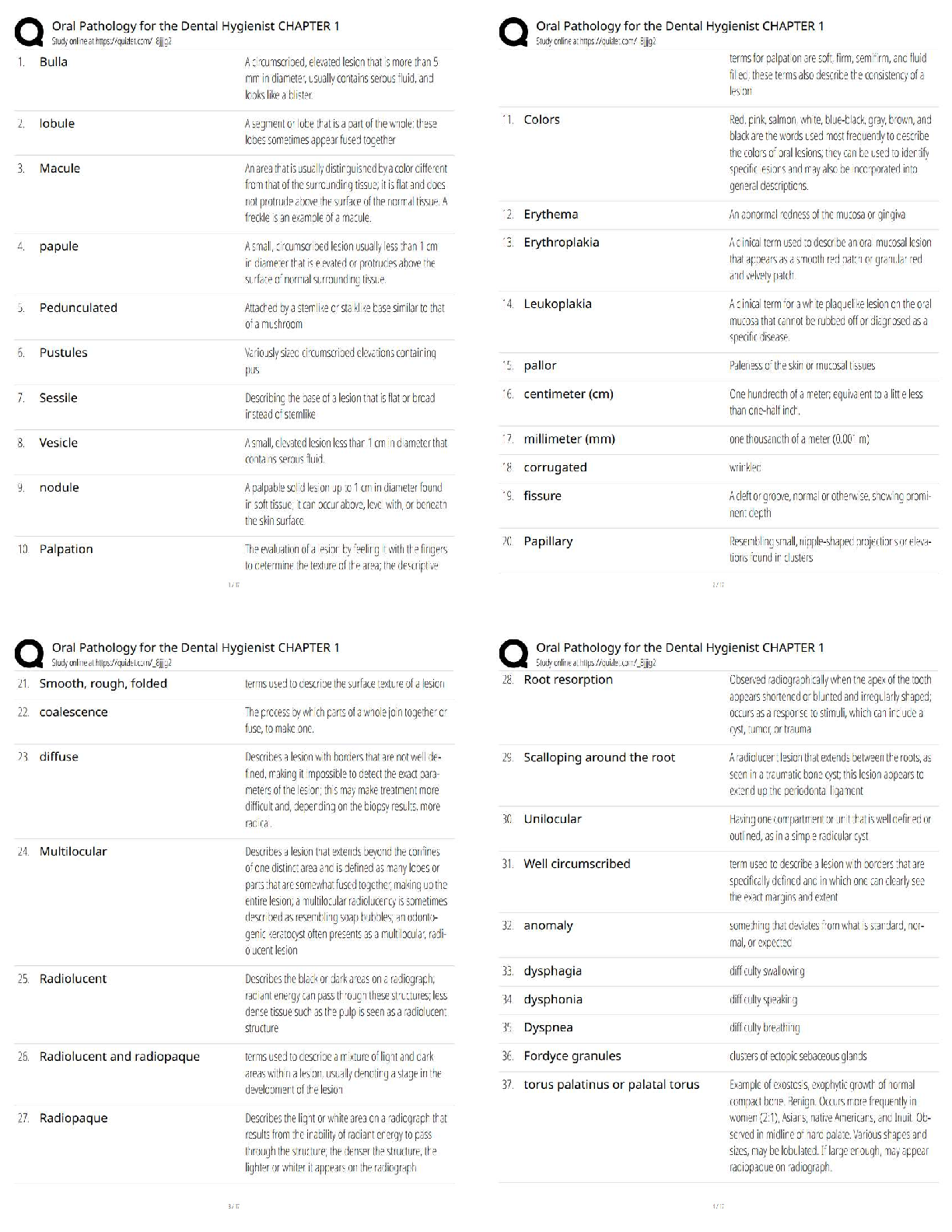
Certified Paraoptometric Anatomy Questions and Answers Rated A Anterior Chamber ✔✔behind the cornea, in front of the iris. Filled with clear watery fluid Aqueous Humor ✔✔clear watery fluid ... produced by ciliary body. provides nutrients for lens and posterior cornea, carries away waste. maintains ocular pressure Choroid ✔✔blood vessels that nourish the retina. between the sclera and retina Ciliary Muscle ✔✔muscle that alters the shape of crystalline lens. Has direct control of focusing ability Conjunctiva ✔✔clear, cellophane like tissue covers sclera and inside surface of lids. Palpebral Conjunctiva ✔✔lines lids Bulbar Conjunctiva ✔✔covers sclera Cornea ✔✔clear, transparent tissue on very front of eye. most powerful refractive media. provides ability to focus on light. Avascular (has no blood vessels) 5 Layers of Cornea ✔✔Front to Back: 1) Epithelium 2) Bowman's layer 3) Stroma 4) Descenet's membrane 5) Endothelium Limbus ✔✔junction of sclera and cornea Crystalline Lens ✔✔Provides focusing power. the 2nd most powerful refractive medium Medial Rectus ✔✔most powerful extraocular muscle. turns eye toward the nose (adduction) Inferior Rectus ✔✔turns eye downward (depresssion). also adducts. can rotate top of eye toward the temple and bottom of the eye towards the nose (extorsion) Lateral Rectus ✔✔moves eye away from nose (abduction) Superior Rectus ✔✔moves eye upwards (elevation). Also adducts. Rotates top of eye toward nose and bottom of eye toward temple (intorsion) Superior Oblique ✔✔Primary role is intorsion. Also depression and abduction Inferior Oblique ✔✔only extraocular muscle that's origin is at front of orbit. Primary role is extorsion. Also elevation and abductino Fovea Centralis ✔✔In macula (approx. 1.5 mm in diameter) where visual acuity is sharpest. Contains highest number of cones which are responsible for daytime and color vision Fundus ✔✔interior surface of eyeball Iris ✔✔colored part of the eye. hole in the middle is pupil. Iris sphincter and dilator muscles control size of pupil for max visual performance Lens ✔✔resilient, transparent structure that focuses light by changes of curvature of front surface. Directly behind the pupil. Macula ✔✔central part of retina. used for seeing detail. Area is 3-5 mm in diameter. Foveal depression is in center Ocular Adnexa ✔✔adjacent structures including: eyelids, lashes, eyebrows, lacrimal apparatus, tarsal plates, orbit, extraocular muscles, conjunctiva. Optic Disk ✔✔appearance of optic nerve when viewing through pupil. Formed by all meetings of retinal nerve fibers. insensitive to light, corresponds to physiological blind spot. used to determine healthiness of nerve Optic Nerve ✔✔carries impulses from retina to brain. transmits signals from rods and cones to brain Orbit ✔✔bony socket that contains eye Posterior Chamber ✔✔located inside eye, behind iris and in front of lens Pupil ✔✔round hole in center of iris which light passes Retina ✔✔seeing part. lines sclera and is where light coming into eye is focused. images that fall on nerve cell are sent to brain for transmission Photoreceptors ✔✔rods and cones Rods ✔✔used for black/white vision. most numerous (120 millions). most sensitive-->night and peripheral vision, motion detection Cones ✔✔used for color vision. only 6-7 million. clear central vision Sclera ✔✔white portion of eye made of tough fibrous tissue. gives shape and structure to eye Suspensory Ligaments ✔✔long thin fibers that connect crystalline lens to ring of ciliary muscles Vitreous Humor ✔✔thick, clear jelly fills eye between lens and retina. help keep eye round Lacrimal Duct ✔✔drainage system for tears Lacrimal gland ✔✔gland that supplies most of tears. located superior and temporally to eye and behind orbital rim. Nasolacrimal duct ✔✔drainage system for tears to leave eye. connected to nasal passage, why nose runs when you cry. Orbital Bones ✔✔1) Maxilla 2) Lacrimal 3) Frontal 4) Palatine 5) Zygomatic 6) Sphenoid 7) Ethmoid [Show More]
Last updated: 2 years ago
Preview 1 out of 6 pages
.png)
Buy this document to get the full access instantly
Instant Download Access after purchase
Buy NowInstant download
We Accept:

Also available in bundle (1)
Click Below to Access Bundle(s)
.png)
Certified Paraoptometric (CPO) Bundled Exams Questions and Answers 100% Pass
Certified Paraoptometric (CPO) Bundled Exams Questions and Answers 100% Pass
By Nutmegs 2 years ago
$18
10
Reviews( 0 )
$8.00
Can't find what you want? Try our AI powered Search
Document information
Connected school, study & course
About the document
Uploaded On
Feb 02, 2023
Number of pages
6
Written in
All

Additional information
This document has been written for:
Uploaded
Feb 02, 2023
Downloads
0
Views
135


.png)