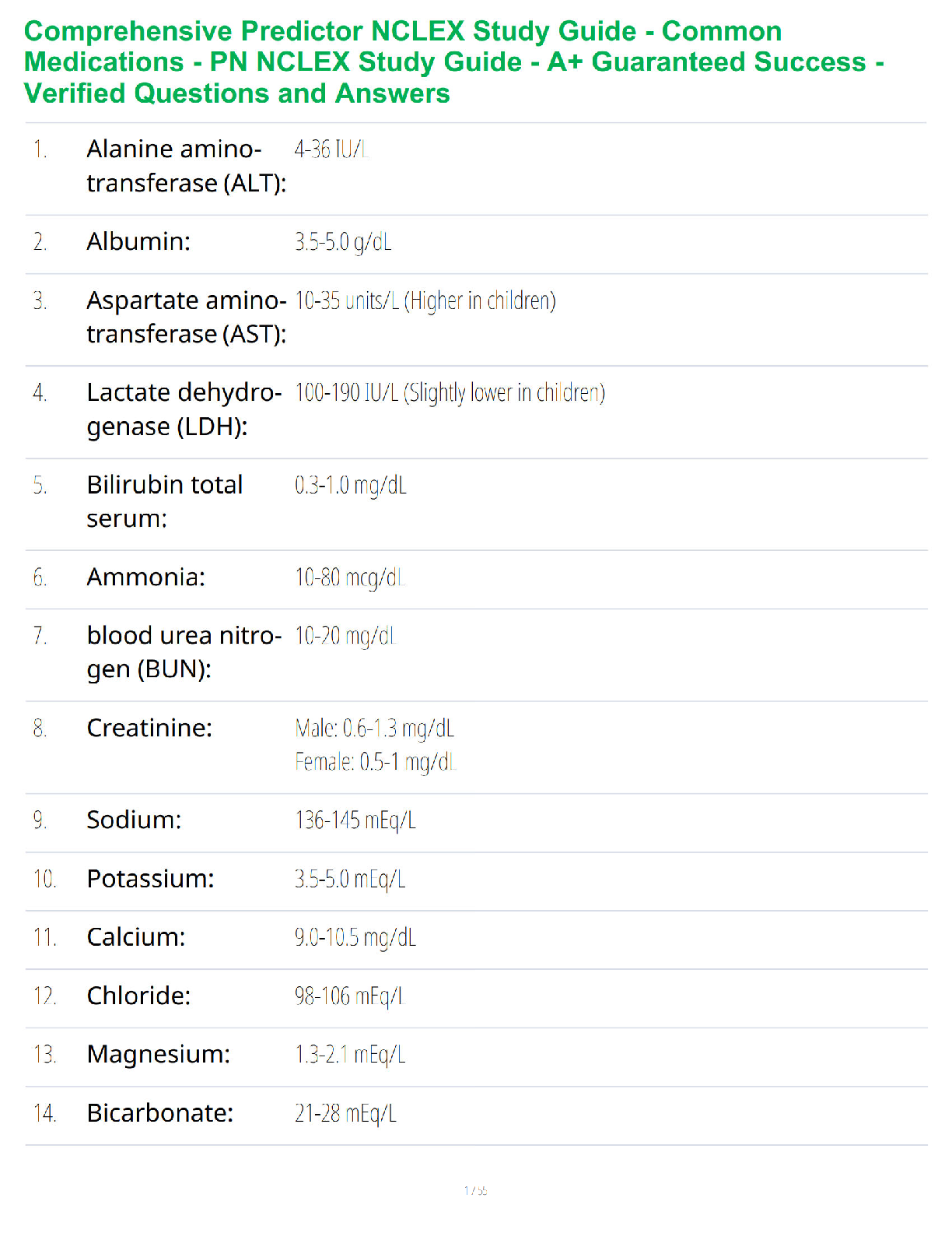
STRUCTURE AND FUNCTION OF THE CV AND LYMPHATIC SYSTEMS STRUCTURE OF
BLOOD VESSELS
Arterial vessels
o Elastic arteries: Contain more elastic fibers than smooth muscle fibers;
absorb energy and stretch.
o Muscular a
...
STRUCTURE AND FUNCTION OF THE CV AND LYMPHATIC SYSTEMS STRUCTURE OF
BLOOD VESSELS
Arterial vessels
o Elastic arteries: Contain more elastic fibers than smooth muscle fibers;
absorb energy and stretch.
o Muscular arteries: Contain fewer elastic fibers and more muscle fibers; can
contract (vasoconstriction) and relax (vasodilation).
Veins
o Thin walled and fibrous with a large diameter
o More numerous than arteries.
o Do not recoil after distention as quickly as arteries.
o Some contain valves.
o Muscle pump: Pushes blood back to the heart.
Capillaries
o Substances move through
Junctions between endothelial cells
Fenestrations (oval windows or pores)
Vesicles moved by active transport
Diffusion
o Blood flow into the capillary beds
Controlled by the contraction and relaxation of smooth muscle bands
(precapillary sphincters) at the junctions between metarterioles and
capillaries
Endothelium roles
o Transportation of substances
o Coagulation
o Antithrombogenesis and fibrinolysis
o Immune system function
o Tissue growth and wound healing
o Vasomotion: Contraction and relaxation of vessels
o Performance of these vital functions through synthesis and the release of
vasoactive chemicals
HEART
Mediastinum
o Area where the heart is located
o Area above the diaphragm and between the lungs
Heart wall
o Epicardium: Outer smooth layer
o Myocardium: Thickest layer of cardiac muscleo Endocardium: Innermost layer
Pericardium - where the heart is enclosed in
o Acts as physical barrier to prevent against infection, inflammation, etc.
o Contains pain receptors – help to elicit reflect changes (BP and HR)
o Double-walled membranous sac
o Parietal: Surface layer
o Visceral: Inner layer; also called epicardium
Pericardial cavity
o Space between the parietal and visceral layers
o Contains pericardial fluid (20 ml)
CHAMBERS OF THE HEART
Right atrium
Left atrium
Atria are separated by the interatrial septum
Right ventricle
Left ventricle
Are separated by the interventricular septum
Thickness of each chamber depends on the pressure or resistance it must overcome
to eject blood
GREAT VESSELS
Superior and inferior venae cavae
o Bring deoxygenated blood from the systemic circulation to the right atrium.
Right and left pulmonary arteries
o Transport unoxygenated blood from the right heart to the right and left lungs.
o Branch into pulmonary capillaries.
Pulmonary veins
o Carry oxygenated blood from the lungs to the left side of the heart.
Aorta
o Delivers oxygenated blood to systemic vessels that supply the body.
CARDIAC CYCLE
Cardiac cycle
o One contraction and one relaxation
o Makes up one heartbeat
Diastole
o Relaxation: Ventricles fill
Systole
o Contraction: Blood leaves the ventricles
Phases of the cardiac cycleo Phase 1: Atrial systole or ventricular diastole
o Phase 2: Isovolumetric ventricular systole
Ventricles build up enough pressure to open semilunar valves against
the pressures of the aorta in the pulmonary artery. Enough pressure
has to build in the ventricles to open the valves
o Phase 3: Ventricular ejection (semilunar valves open)
o Phase 4: Isovolumetric ventricular relaxation (aortic valve closes)
o Phase 5: Passive ventricular filling (mitral and tricuspid
o valves open)
BLOOD FLOW
Unoxygenated (venous) blood from systemic circulation enters the right atrium
through the superior and inferior venae cavae.
From the atrium, the blood passes through the right AV (tricuspid) valve into the
right ventricle.
In the ventricle, the blood flows from the inflow tract to the outflow tract and then
through the pulmonic semilunar valve (pulmonary valve) into the pulmonary artery,
which delivers it to lungs for oxygenation
Oxygenated blood from the lungs enters the left atrium through the four pulmonary
veins (two from the left lung and two from the right).
[Show More]


, 100% Correct, Download to Score A.png)